
Retroperitoneal fibrosis, also known as Ormond’s disease, is a rare condition characterized by the development of excess fibrous tissue in the retroperitoneal space, the area behind the abdominal cavity. This fibrous tissue can encase and obstruct structures such as the ureters, leading to complications like kidney damage. The exact cse of the disease is often unknown.
Symptoms often include abdominal pain, back pain, and renal impairment. Diagnosis typically involves imaging studies, and treatment may include medications, or surgery.
What is retroperitoneum?
The retroperitoneum, or retroperitoneal space, is the anatomical area located behind (posterior to) the peritoneum, the lining of the abdominal cavity. It is a potential space that contains various organs and structures that are not enclosed by the peritoneal sac.
Organs and structures in the retroperitoneum
- Kidneys
- Ureters
- Adrenal Glands
- Pancreas (except for the tail): The pancreas is involved in digestion and blood sugar regulation. Most of the pancreas lies in the retroperitoneal space.
- Duodenum (except for the first part): This is the first section of the small intestine, where initial digestion occurs.
- Ascending and Descending Colon: Parts of the large intestine that absorb water and transport waste material.
- Aorta and Inferior Vena Cava
- Lymph Nodes and Vessels
- Sympathetic Nerves and Plexuses
What is retroperitoneal fibrosis (Ormond’s disease)?
Retroperitoneal fibrosis, also known as Ormond’s disease, is a rare disorder characterized by the development of excess fibrous tissue in the retroperitoneal space, the area behind the peritoneum in the abdomen. This fibrotic tissue can encase and compress the organs and structures in the retroperitoneum, most notably the ureters, which are the tubes that carry urine from the kidneys to the bladder.
Risk factors
Retroperitoneal fibrosis is more common in males than females, with a male-to-female ratio of approximately 2:1 to 3:1. Retroperitoneal fibrosis is a rare disease, with an estimated incidence of 0.1 to 1.3 cases per 100,000 people per year. Several risk factors and causes have been associated with its development:
- Age: The condition is most commonly diagnosed in middle-aged individuals, typically between 40 and 60 years old.
- Gender: As noted, males are at a higher risk than females.
- Autoimmune Reaction: Many cases of retroperitoneal fibrosis are thought to be due to an autoimmune reaction, where the body’s immune system mistakenly attacks its own tissues, leading to inflammation and fibrosis.
- Medications: Certain drugs have been linked to the development of retroperitoneal fibrosis, including:
- Ergot derivatives: Used for migraine treatment.
- Beta-blockers: Such as propranolol, used to treat high blood pressure and heart conditions.
- Methyldopa: An antihypertensive drug.
- Infections: Infections, particularly chronic infections, can trigger inflammation leading to fibrosis. This includes infections such as tuberculosis and certain fungal infections.
- Malignancies: Some cancers, particularly those in nearby structures such as lymphoma, can cause secondary retroperitoneal fibrosis.
- IgG4-Related Disease: This is an immune-mediated condition that can cause fibrosis in various organs, including the retroperitoneum.
- Surgical or Traumatic Injury: Previous abdominal surgeries or trauma to the retroperitoneal area can lead to fibrotic reactions.
- Idiopathic: In many cases, no specific cause can be identified, and the condition is termed idiopathic retroperitoneal fibrosis.
Symptoms
The symptoms of retroperitoneal fibrosis (Ormond’s disease) can vary depending on the extent and location of the fibrotic tissue and the organs affected. Early symptoms are often nonspecific and can be mistaken for other conditions, but as the disease progresses, more specific symptoms may develop due to the compression of retroperitoneal structures. These include:
- Abdominal Pain: Persistent or intermittent pain in the lower abdomen or flank region is one of the most common symptoms. The pain can range from mild to severe.
- Back Pain: Pain may also occur in the lower back due to the involvement of the lumbar region.
- Renal Symptoms: As the fibrous tissue often encases the ureters, obstructing urine flow, symptoms related to kidney function can occur, including:
- Reduced urine output
- Hematuria (blood in urine)
- Renal colic (severe pain due to kidney stones)
- Hypertension (high blood pressure) due to kidney impairment
- Constitutional Symptoms: These are general symptoms that can affect the entire body, such as:
- Fatigue and weakness
- Unintended weight loss
- Fever
- Lower Limb Swelling: If the inferior vena cava is compressed, it can lead to swelling (edema) in the legs.
- Gastrointestinal Symptoms: Nausea, vomiting, and changes in bowel habits may occur if the gastrointestinal tract is affected.
- Hydronephrosis: This is a condition characterized by swelling of the kidneys due to urine build-up, often detected through imaging studies.
- Advanced Symptoms: In advanced cases where significant obstruction occurs, patients may experience severe kidney dysfunction, electrolyte imbalances, and complications related to chronic kidney disease.
Diagnosis
Diagnosing retroperitoneal fibrosis (Ormond’s disease) involves a combination of clinical evaluation, imaging studies, and sometimes laboratory tests and biopsy to confirm the presence of fibrous tissue and rule out other conditions. Here’s an overview of the diagnostic process:
- Medical History and Physical Examination: The doctor will assess the patient’s symptoms, medical history, and perform a physical examination to look for signs such as abdominal or flank tenderness and signs of kidney dysfunction.
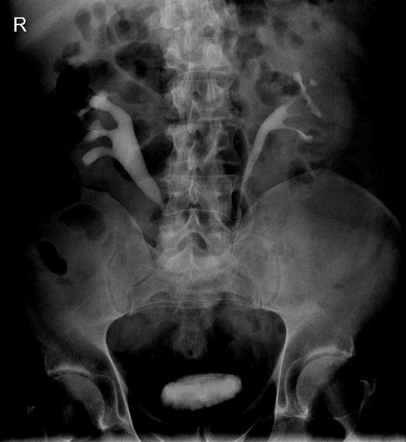
- Computed Tomography (CT) Scan: A CT scan of the abdomen and pelvis provides detailed images of the retroperitoneal space, helping to identify the fibrotic mass and any effects on the kidneys, ureters, and other structures. CT can show the extent of fibrosis and any resulting hydronephrosis (swelling of the kidneys).
- Magnetic Resonance Imaging (MRI): MRI offers excellent soft tissue contrast and can be used to distinguish between fibrous tissue and other types of masses, such as tumors. It is helpful in evaluating the involvement of adjacent organs and blood vessels.
- Ultrasound: While not as detailed as CT or MRI, an ultrasound can be used to assess kidney size and detect hydronephrosis.
- Blood Tests: To evaluate kidney function (e.g., creatinine levels), assess inflammatory markers, and rule out other conditions.
- Urinalysis: To check for hematuria (blood in urine) and other signs of kidney impairment.
- Tissue Biopsy: In some cases, a biopsy of the fibrous tissue may be performed to rule out malignancy and confirm the diagnosis of idiopathic retroperitoneal fibrosis. This involves obtaining a tissue sample through needle biopsy or surgical exploration.
Differential diagnosis
The diagnostic process may also involve ruling out other conditions that can mimic retroperitoneal fibrosis, such as:
- Malignancies (e.g., lymphoma, sarcoma)
- Infectious diseases (e.g., tuberculosis)
- Other causes of ureteral obstruction
Complications
Retroperitoneal fibrosis (Ormond’s disease) can lead to several urologic complications due to the fibrous tissue encasing and compressing the ureters and other structures in the retroperitoneal space. Here are the primary urologic complications associated with this condition:
- Ureteral Obstruction
- Hydronephrosis
- Renal Insufficiency
- Acute Renal Failure
- Urinary Tract Infections (UTIs)
- Renal Colic
- Hypertension
- Electrolyte Imbalances
Treatment
The treatment of retroperitoneal fibrosis (Ormond’s disease) aims to reduce inflammation, manage symptoms, and prevent complications such as kidney damage. Treatment typically involves a combination of medications and, in some cases, surgical interventions.
Medical Treatment
- Corticosteroids: Corticosteroids, such as prednisone, are often the first-line treatment to reduce inflammation and shrink the fibrotic tissue. The dosage is usually high initially, then gradually tapered over several months as the condition improves.
- Immunosuppressive Medications: In cases where corticosteroids are not sufficient or cannot be used long-term, additional immunosuppressive drugs such as azathioprine, methotrexate, or mycophenolate mofetil may be prescribed. These medications help control the immune response and prevent further fibrosis.
- Tamoxifen: This anti-estrogen medication has been used in some cases for its anti-fibrotic properties, although its exact mechanism in retroperitoneal fibrosis is not fully understood.
- Biologic Agents: Biologics such as rituximab or infliximab may be considered for patients with refractory disease or those with an IgG4-related disease component.
Surgical Treatment
- Ureterolysis: A surgical procedure to free the ureters from fibrous tissue and relieve obstruction. It may involve wrapping the ureters with omental tissue to prevent re-encasement.
- Ureteral Stenting or Nephrostomy: Placement of stents in the ureters or nephrostomy tubes in the kidneys can temporarily relieve obstruction and preserve kidney function while other treatments take effect.
- Laparoscopic or Open Surgery: In severe cases, more invasive surgery may be needed to remove the fibrotic tissue or bypass obstructed areas.
Prognosis
The prognosis of retroperitoneal fibrosis (Ormond’s disease) depends on several factors, including the severity of the condition, the effectiveness of treatment, and the presence of any underlying causes or complications. Here’s a summary of key aspects of the prognosis:
General Prognosis
- Early Diagnosis and Treatment: The prognosis is generally better with early diagnosis and prompt treatment. Early intervention can help prevent significant kidney damage and other complications. Effective management of inflammation and obstruction usually leads to improved outcomes and symptom relief.
- Response to Treatment: Many patients respond well to corticosteroids and other immunosuppressive therapies, leading to a reduction in fibrotic tissue and improvement in symptoms. In cases with good control of inflammation and successful relief of ureteral obstruction, kidney function can be preserved or restored.
- Chronicity and Recurrence: Some patients may experience recurrence of symptoms or disease progression, especially if the underlying cause is not addressed. Long-term monitoring is often required to manage potential recurrences and adjust treatment as needed.
- Complications: The presence of complications such as severe kidney damage, chronic renal insufficiency, or recurrent urinary tract infections can affect the overall prognosis. Timely management of these complications is crucial to improving long-term outcomes.
- Underlying Causes: If retroperitoneal fibrosis is secondary to an underlying condition (such as malignancy or autoimmune disease), the prognosis may be influenced by the management of that primary condition. Idiopathic cases, where no clear cause is identified, may have a more variable prognosis depending on how well the condition responds to treatment.
Long-Term Outlook
- Kidney Function: Many patients can achieve good control of their symptoms and maintain kidney function with appropriate treatment. However, those with significant or prolonged obstruction may experience some degree of lasting kidney impairment.
- Quality of Life: With effective treatment, most patients can experience significant relief from symptoms and improvements in quality of life.
- Follow-Up Care: Regular follow-up with imaging and laboratory tests is essential to monitor the condition, adjust treatment, and detect any recurrence or complications early.
Summary
Retroperitoneal fibrosis is a rare condition characterized by the formation of excess fibrous tissue in the retroperitoneal space, located behind the peritoneum in the abdomen. This fibrotic tissue can encase and compress organs, particularly the ureters, leading to obstruction and potential kidney damage. Management of retroperitoneal fibrosis requires a multidisciplinary approach to address the complex and potentially chronic nature of the disease.
Prof. Dr. Emin ÖZBEK
Urologist
Istanbul- TURKIYE

Leave a Reply