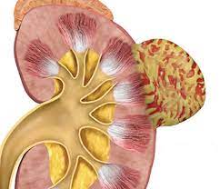
Renal angiomyolipoma (AML) is a benign kidney tumor composed of three types of tissues: blood vessels, muscle, and fat. It is the most common benign renal tumor, often found incidentally during imaging for other conditions. Though typically asymptomatic and non-cancerous, larger AMLs can cause symptoms like pain, blood in the urine (hematuria), or kidney function impairment due to bleeding or pressure effects. These tumors are frequently associated with a genetic condition called tuberous sclerosis complex (TSC), but they can also occur sporadically. Treatment depends on size and symptoms, ranging from observation to surgical or minimally invasive interventions.:
What is renal angiomyolipoma?

Renal angiomyolipoma is a non-cancerous (benign) tumor that develops in the kidneys. It is made up of a mixture of blood vessels, smooth muscle cells, and fat tissue. AMLs are the most common type of benign kidney tumors and can occur sporadically or in association with a genetic disorder called TSC, which can lead to multiple tumors in both kidneys.
Most AMLs are asymptomatic and are often discovered incidentally during imaging tests like ultrasounds or CT scans done for other reasons. However, larger AMLs can sometimes cause symptoms such as flank pain, blood in the urine (hematuria), or even bleeding if a rupture occurs. Depending on the size and symptoms, treatment can include monitoring, medication, embolization (blocking blood supply to the tumor), or surgical removal.
Incidence of AML
Overall, sporadic (non-TSC-related) AMLs are more common in women and are typically diagnosed between the ages of 40 and 60. The incidence of renal angiomyolipomavaries depending on the population:
- In the general population, the incidence is estimated to be around 0.1% to 0.3%.
- Among individuals with TSC, the incidence is much higher, occurring in up to 80% of patients.
- In patients with LAM, about 30% to 50% may develop renal AMLs.
Clasification of AML
AML can be classified into several types based on their association with genetic conditions and histological characteristics:
- Sporadic AML: Occurs without any underlying genetic condition. Typically presents as isolated tumors and is more common in women, especially those aged 40 to 60.
- Hereditary AML: Associated with genetic disorders, primarily: TS) and LAM.
- Histological Variants: Classic AML, Epithelioid AML, Pleomorphic AML:
- Giant AML: Refers to very large AMLs, often exceeding 10 cm in size.
Diseases associated with AML
Renal Angiomyolipoma is primarily associated with:
- Tuberous sclerosis complex (TSC)
- Lymphangioleiomyomatosis (LAM)
- Neurofibromatosis type 1
- Von Hippel-Lindau disease
Causes of AML
The causes of AML can be grouped into two main categories:
- Sporadic (Isolated) AML: Occurs without any underlying genetic condition.
- Genetic-Associated AML: In this cathegoria there are 2 diseases: Tuberous sclerosis complex (TSC): A genetic disorder caused by mutations in the TSC1 or TSC2 genes, leading to the development of multiple benign tumors in various organs, including the kidneys. Lymphangioleiomyomatosis (LAM): A rare disease that primarily affects women and is linked to mutations in the TSC2 gene, often leading to the development of renal AMLs.
Symptoms of AML
Symptoms of AML can vary depending on the size and number of tumors. Many AMLs are asymptomatic, especially when small. However, larger or multiple tumors may cause noticeable symptoms, including:
- Flank or abdominal pain
- Hematuria (blood in the urine)
- Palpable mass
- Hypertension
- Anemia
- Retroperitoneal hemorrhage (wunderlich syndrome): a rare but serious condition where the tumor ruptures, leading to sudden and severe abdominal or flank pain, dizziness, and a drop in blood pressure.
Diagnosis of AML
The diagnosis of AML primarily involves imaging techniques that can identify the tumor’s unique composition of fat, muscle, and blood vessels. Diagnostic steps include:
- Ultrasound: Common initial imaging test. AMLs appear as well-defined, hyperechoic (bright) masses due to their fat content.
- Computed Tomography (CT) Scan: AMLs typically show a characteristic fat component, making them easily distinguishable from other kidney masses.
- Magnetic Resonance Imaging (MRI): Effective in differentiating AML from other renal tumors, especially in cases where the fat content is low.
- Angiography: Occasionally used to visualize the blood supply of the tumor. Helps in planning treatment, such as embolization.
- Biopsy: Rarely required since imaging usually provides a clear diagnosis. May be considered if imaging findings are ambiguous and malignancy cannot be ruled out.
Treatment of renal angiomyolipoma
The treatment of AML depends on the size of the tumor, symptoms, and potential risk factors, such as bleeding. Common treatment options include:
- Active Surveillance: For small, asymptomatic AMLs (usually less than 4 cm). Regular imaging follow-ups (e.g., ultrasound or CT scan) to monitor tumor growth or changes.
- Medications: mTOR Inhibitors (e.g., Everolimus) used to shrink tumors, especially in cases associated with TSC or LAM.
- Embolization: A minimally invasive procedure to block the blood supply to the tumor. Effective for controlling bleeding or reducing the size of larger AMLs. Often used as a first-line treatment for symptomatic AMLs.
- Percutaneous Ablation: Techniques like radiofrequency ablation or cryotherapy to destroy the tumor using heat or cold.
- Surgical Intervention: Partial Nephrectomy or total nephrectomy
- Emergency Management: For cases of sudden bleeding embolization or surgery may be performed to stop the bleeding and stabilize the patient.
Indications of surgery for AML
Surgery for AML is typically indicated in the following scenarios:
- Large tumors (≥ 4 cm):
- Symptomatic tumors:
- Risk of hemorrhage:
- Rapid tumor growth:
- Suspicion of malignancy:
- Failure of minimally ınvasive treatments:
- Tumor in high-risk locations:
Preventive measures for AML
Preventing renal angiomyolipoma can be challenging since most cases occur sporadically or are linked to genetic conditions like TSC and LAM. However, some measures may help in early detection and management to reduce the risk of complications:
- Genetic Counseling
- Regular Monitoring
- Medication Management (mTOR inhibitors)
- Avoid High-Risk Activities
- Blood Pressure Control
Prognosis
AML is a benign tumor. It is not cancerous and does not metastasize. In sporadic cases and tumors smaller than 4 cm, the disease course is generally good. However, in larger tumors and those associated with genetic diseases such as Tuberous TSC and LAM, the prognosis should be monitored more closely compared to sporadic cases.
Summary
renal angiomyolipoma is a benign kidney tumor characterized by a mixture of blood vessels, smooth muscle, and fat. It is the most common type of benign renal tumor, often discovered incidentally during imaging. While many AMLs are asymptomatic, larger tumors can cause symptoms such as flank pain and hematuria. The condition is commonly associated with genetic disorders like TSC and LAM. Diagnosis is primarily through imaging techniques such as ultrasound and CT scans. Treatment options range from active surveillance for small, asymptomatic tumors to surgical intervention for larger or symptomatic cases. The overall prognosis is favorable, especially with appropriate monitoring and management.
Prof. Dr. Emin ÖZBEK
Urologist
Istanbul- TURKIYE


Leave a Reply