
Uhe ureters serve as vital conduits for the transportation of urine from the kidneys to the urinary bladder, facilitating the elimination of waste from the body. Understanding their anatomy and function is essential for diagnosing and treating various urinary system disorders and conditions.
Here i will give general informations about ureters.
What is “ureter”?
The ureter is a muscular tube in the urinary system of mammals, including humans. It serves to transport urine from the kidneys to the urinary bladder. Each human typically has two ureters, one connecting each kidney to the bladder. The ureters are responsible for facilitating the flow of urine by utilizing peristaltic contractions, which are rhythmic waves of muscular movements. These contractions help propel urine from the kidneys to the bladder, where it is stored until it is expelled from the body during urination.
Anatomy of “ureter”
The ureter is a tubular structure that connects the kidneys to the urinary bladder in the urinary system. It’s an essential part of the urinary tract, responsible for transporting urine from the kidneys, where it’s produced, to the bladder, where it’s stored until it’s expelled from the body during urination. Here’s a detailed anatomy of the ureter:
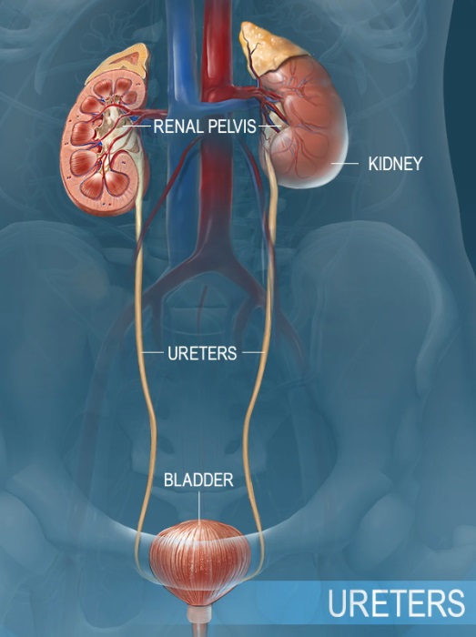
- Location: Ureters are retroperitoneal structures, meaning they lie behind the peritoneum—the membrane lining the abdominal cavity. They run from the renal pelvis of each kidney to the urinary bladder.
- Length: The length of the ureters can vary, but on average, they are about 25 to 30 centimeters long.
- Structure: The ureter is a muscular tube with three layers:
- Mucosa: The innermost layer, consisting of transitional epithelium, which allows the ureter to stretch to accommodate urine flow without damage.
- Muscularis: The middle layer, composed of smooth muscle fibers arranged in longitudinal and circular layers. These muscles contract in a coordinated manner to propel urine through the ureter via peristaltic movements.
- Adventitia: The outermost layer, made up of connective tissue, which anchors the ureter to surrounding structures.
- Blood Supply: Ureters receive their blood supply from branches of the renal, gonadal, and common iliac arteries.
- Nerve Supply: Nerves from the autonomic nervous system innervate the ureter, controlling its peristaltic contractions and sensation.
- Course:
- Renal Pelvis: The ureter begins at the renal pelvis, a widened funnel-shaped structure that collects urine from the kidney’s collecting ducts.
- Abdomen: From the renal pelvis, the ureter descends downward behind the peritoneum, running along the posterior abdominal wall and crossing over the pelvic brim into the pelvic cavity.
- Pelvis: Within the pelvis, the ureter travels along the sidewall, crossing the pelvic inlet and entering the urinary bladder from below.
- Ureterovesical Junction (UVJ): The point where the ureter meets the bladder is called the ureterovesical junction. It features a one-way valve mechanism that prevents urine reflux from the bladder back into the ureter.
- Ureteral Peristalsis: Peristaltic contractions of the ureter walls help propel urine from the kidneys to the bladder. This mechanism ensures the unidirectional flow of urine.
Narrow areas of the ureters and clinical significance
The ureters have several narrow areas that are clinically significant because they are common sites for obstruction or kidney stone passage. The three primary narrowings are:
- Ureteropelvic Junction (UPJ): This is the area where the ureter meets the renal pelvis. It’s a common site for obstruction, which can cause hydronephrosis (swelling of the kidney due to urine buildup).
- Pelvic Ureteric Junction (PUJ): This is where the ureter crosses over the pelvic brim. Stones or strictures in this area can cause similar symptoms and complications as those seen at the UPJ.
- Ureterovesical Junction (UVJ): This is where the ureter connects to the bladder. This junction can be a site of obstruction or infection and is often where stones get stuck before entering the bladder.
These narrow areas are significant in clinical practice because blockages here can lead to severe pain, infection, and kidney damage if not treated promptly.
Functions of ureters
The ureter plays several crucial functions in the urinary system. Here are its primary roles:
- Transportation of Urine: The main function of the ureter is to transport urine from the kidneys to the urinary bladder. Urine is continuously produced by the kidneys as they filter waste products and excess substances from the blood. The ureter acts as a conduit, facilitating the passage of urine from the renal pelvis of each kidney to the bladder.
- Peristalsis: The walls of the ureter contain smooth muscle tissue that undergoes rhythmic contractions known as peristalsis. These contractions propel urine downward through the ureter toward the bladder. Peristalsis helps to overcome gravity and move urine efficiently, even when the body is in various positions.
- One-Way Valve Mechanism: The ureterovesical junction (UVJ), where the ureter enters the bladder, features a one-way valve mechanism. This valve prevents urine from flowing backward from the bladder into the ureter, thereby maintaining the unidirectional flow of urine from the kidneys to the bladder.
- Prevention of Urinary Tract Infections: The continuous flow of urine through the ureters helps flush out bacteria and other harmful substances from the urinary tract. This helps to prevent the buildup of pathogens in the ureters and reduces the risk of urinary tract infections (UTIs).
- Maintenance of Urine Composition: While the primary function of the ureter is transportation, it also contributes to the maintenance of urine composition. The selective reabsorption and secretion of ions, water, and other substances by the renal tubules in the kidneys influence the composition of urine that ultimately reaches the bladder via the ureters.
- Sensory and Reflex Functions: The ureter contains sensory nerves that detect the presence of urine and stretch within its walls. These sensory signals trigger reflex responses that regulate the rate and intensity of ureteral peristalsis, ensuring efficient urine transport.
Common disorders of ureters
The ureters can be affected by various disorders that can impair their function and lead to complications. Some common disorders of the ureters include:
- Ureteral Obstruction: This occurs when there is a blockage in one or both ureters, preventing the normal flow of urine from the kidneys to the bladder. Causes of obstruction can include kidney stones, blood clots, tumors, ureteral strictures (narrowing), or external compression from neighboring structures. Ureteral obstruction can lead to pain, urinary tract infections, kidney damage, and impaired kidney function.
- Ureteral Stricture: Narrowing of the ureter, known as ureteral stricture, can result from scarring due to inflammation, previous surgeries, radiation therapy, or congenital abnormalities. Ureteral strictures can impede urine flow, leading to hydronephrosis (fluid buildup in the kidney), recurrent urinary tract infections, and kidney damage.
- Ureteral Reflux: Ureteral reflux, also called vesicoureteral reflux (VUR), occurs when urine flows backward from the bladder into one or both ureters. This condition is more common in children and can predispose individuals to urinary tract infections and kidney damage if left untreated. Ureteral reflux may be congenital or acquired and can result from abnormalities in the ureterovesical junction (UVJ) or bladder dysfunction.
- Ureteral Injury: Trauma, surgical procedures, or medical interventions in the pelvic or abdominal region can lead to ureteral injury. Ureteral injuries may involve lacerations, tears, or complete transections of the ureter, leading to leakage of urine into the abdominal cavity, infection, and subsequent complications. Prompt diagnosis and surgical repair are essential to prevent long-term complications.
- Ureteral Calculi (Kidney Stones): Kidney stones can form within the kidney and may migrate into the ureter, causing obstruction and severe pain. The passage of kidney stones through the ureter can result in ureteral colic, characterized by intense flank pain that radiates to the groin. Other symptoms may include hematuria (blood in the urine), urinary urgency, and nausea. Treatment may involve pain management, hydration, and interventions to facilitate stone passage or surgical removal.
- Ureteral Tumors: Tumors can develop within the ureter, although they are relatively rare compared to tumors in other parts of the urinary system. Ureteral tumors may be benign or malignant (cancerous) and can obstruct urine flow, leading to symptoms such as pain, hematuria, and urinary tract infections. Treatment typically involves surgical resection of the tumor, sometimes followed by chemotherapy or radiation therapy for malignant tumors.
Congenital anomalies of ureters
Congenital anomalies of the ureters are structural abnormalities that are present at birth. These anomalies can affect the shape, size, position, or function of the ureters and may lead to various urinary system complications. Some common congenital anomalies of the ureters include:
- Ureteropelvic Junction Obstruction (UPJO): UPJO is a condition where there is a blockage or narrowing at the junction where the ureter connects to the renal pelvis (ureteropelvic junction). This obstruction impedes the flow of urine from the kidney to the ureter, leading to hydronephrosis (dilation of the renal pelvis and calyces due to urine buildup). UPJO can cause flank pain, urinary tract infections, and if severe and untreated, kidney damage.
- Duplex Collecting System: In this anomaly, there is a duplication of the ureter and renal pelvis in one or both kidneys. This results in two ureters draining from a single kidney or a portion of the kidney. Duplex collecting systems can be complete (with two separate ureters draining into the bladder) or incomplete (with one ureter draining into the other ureter or elsewhere in the urinary tract). Duplex collecting systems may increase the risk of urinary tract infections and other complications.
- Ureteral Duplication: Ureteral duplication is a condition where a single kidney has two ureters draining separately into the bladder. Each ureter may have its own opening in the bladder (complete duplication) or share a common opening (partial duplication). Ureteral duplication can predispose individuals to urinary reflux, urinary tract infections, and kidney stones.
- Ectopic Ureter: An ectopic ureter is a congenital anomaly where the ureter terminates at an abnormal location, usually outside the bladder. In males, an ectopic ureter may connect to the urethra, seminal vesicles, or other structures in the genitourinary system. In females, it may connect to the urethra, vagina, uterus, or other structures. Ectopic ureters can lead to urinary incontinence, recurrent urinary tract infections, and kidney damage.
- Ureterocele: A ureterocele is a cystic dilation or pouching of the distal end of the ureter near its insertion into the bladder. Ureteroceles may obstruct urine flow, leading to urinary retention, urinary tract infections, and kidney damage. They can be associated with duplex collecting systems or other congenital anomalies.
- Ureteral Atresia: Ureteral atresia is a rare congenital anomaly characterized by the complete absence or closure of a segment of the ureter. This anomaly obstructs urine flow and may lead to hydronephrosis and kidney dysfunction.
- Retrocaval ureter: A “retrocaval ureter” is a rare anatomical anomaly where the ureter passes behind the inferior vena cava instead of in front of it. This can lead to obstruction or compression of the ureter, potentially causing symptoms like pain or urinary issues.
Summary
The ureter is a muscular tube in the urinary system that connects each kidney to the urinary bladder. Its primary function is to transport urine from the kidneys, where it’s produced, to the bladder, where it’s stored until expelled from the body during urination. The ureter employs peristaltic contractions to propel urine downward, overcoming gravity. Common disorders of the ureters include obstruction, stricture, reflux, injury, calculi, and tumors. Congenital anomalies, such as UPJO, duplex collecting systems, ureteral duplication, ectopic ureter, ureterocele, and ureteral atresia, can affect its structure and function. Management involves early diagnosis and appropriate treatment to prevent complications and preserve urinary system health.
Prof. Dr. Emin ÖZBEK
Urologist
Istanbul- TURKIYE

Leave a Reply