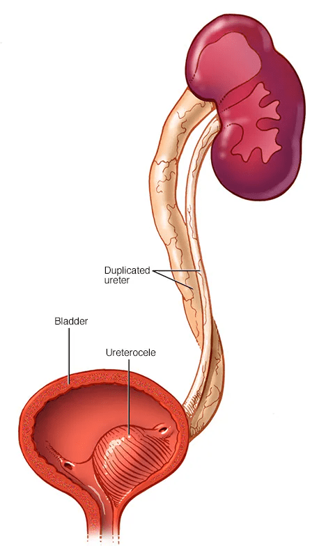
A ureterocele is a congenital (present at birth) condition in which the end of a ureter—the tube that carries urine from the kidney to the bladder—swells and forms a sac-like pouch inside the bladder. This swelling can block the normal flow of urine, leading to urine backup, potential kidney damage, and urinary tract infections (UTIs). Ureteroceles are more common in females and can affect one or both ureters. Treatment varies based on severity and may include surgery to relieve the obstruction and improve urinary drainage.
What is ureterocele?
A ureterocele is a medical condition where the lower part of the ureter—the tube that transports urine from the kidney to the bladder—bulges out or swells at its connection to the bladder, forming a balloon-like pouch. This can partially or fully obstruct urine flow from the kidney to the bladder, leading to issues like urinary retention, UTIs, and sometimes kidney damage due to the backup of urine. Ureteroceles are typically congenital, meaning they are present at birth, and they can affect one or both ureters. They are often diagnosed in children and are more common in females.

Causes of ureterocele
The exact cause of a ureterocele is not fully understood, but it is believed to be related to developmental abnormalities in the urinary tract that occur before birth. Some possible contributing factors include:
- Congenital malformation: Ureteroceles typically develop as a result of a congenital defect during fetal development. This can cause the distal (lower) end of the ureter to narrow, preventing proper urine flow into the bladder and leading to swelling.
- Duplication of the ureter: In some cases, a person may be born with two ureters leading from one kidney (called a duplicated collecting system). The upper ureter in this system is more prone to forming a ureterocele due to abnormal development.
- Obstruction at the ureterovesical junction: This refers to an obstruction where the ureter meets the bladder. A structural defect at this junction can prevent urine from draining normally, resulting in the formation of a ureterocele.
- Genetic factors: While a direct genetic cause hasn’t been clearly identified, congenital urinary tract abnormalities, including ureteroceles, may run in families, indicating a potential genetic predisposition.
Symptoms of ureterocele
Symptoms of a ureterocele can vary depending on its size, severity, and whether it causes an obstruction in the urinary tract. In some cases, especially in children, a ureterocele may not show any symptoms and might be discovered incidentally during imaging tests. Common symptoms include:
- Urinary Tract Infections (UTIs): Frequent UTIs are one of the most common signs, especially in young children.
- Abdominal or Flank Pain: Pain in the lower abdomen or sides (flank area) can result from urinary blockage or infection.
- Difficulty Urinating or Frequent Urination: Some people may experience urinary retention, difficulty starting urination, or an increased urge to urinate.
- Hematuria (Blood in the Urine): Blood in the urine may appear as a result of infection or irritation in the urinary tract.
- Vesicoureteral Reflux: This condition involves the backflow of urine from the bladder into the ureters and kidneys, leading to kidney swelling and damage over time.
- Poor Growth or Failure to Thrive: In infants, chronic infection or kidney problems may affect normal growth and development.
Diagnosis of ureterocele
The choice of diagnostic test(s) depends on the individual’s symptoms and the complexity of the suspected ureterocele. Common diagnostic methods include:
- Ultrasound: It can show an enlarged ureter, a pouch within the bladder, or signs of kidney swelling (hydronephrosis) caused by urine backup.
- Voiding Cystourethrogram (VCUG): This test involves filling the bladder with a contrast dye and taking X-ray images while the patient urinates. It can reveal if urine flows back up the ureters (vesicoureteral reflux), a common complication of ureteroceles.
- Intravenous Pyelogram (IVP): In this test, a contrast dye is injected into a vein and X-ray images are taken as the dye moves through the kidneys, ureters, and bladder. This helps to highlight any abnormalities in the urinary tract, such as a ureterocele or blockage.
- Magnetic Resonance Urography (MRU) or CT Urography: These are particularly useful if there is a complex anatomy, like duplicated ureters.
- Renal Scintigraphy (DMSA Scan): This nuclear medicine scan evaluates kidney function by assessing how well each kidney filters blood. It helps determine if kidney function is compromised by the ureterocele.
- Cystoscopy: It allows doctors to directly visualize the ureterocele and assess its size and position in the bladder.
Complications of ureterocele
If left untreated, a ureterocele can lead to several complications, which may impact kidney and urinary tract health. Common complications include:
- Urinary tract ınfections
- Vesicoureteral reflux
- Kidney damage
- Urinary retention and obstruction
- Poor growth and development
- Bladder dysfunction
Treatment of ureterocele
Treatment of a ureterocele depends on its size, location, the severity of symptoms, and whether there is a risk of kidney damage. The main goal is to restore normal urine flow, prevent infections, and protect kidney function. Here are common treatment options:
- Endoscopic Surgery: This minimally invasive procedure involves the puncture or incise the ureterocele. This allows urine to flow freely from the ureter to the bladder and can reduce the risk of recurrent infections and obstruction.
- Ureteral Reimplantation: In cases where the ureterocele or ureteral anatomy is complex, a surgical reimplantation may be needed. This involves repositioning the ureter’s connection to the bladder to ensure proper urine flow and prevent reflux. This procedure is more invasive and often done when simpler treatments are not sufficient.
- Upper Pole Nephrectomy: If the ureterocele is associated with a duplicated ureter system (two ureters on one side) and has significantly damaged the upper portion of the kidney, removing the affected portion may be necessary. This is done to prevent ongoing complications and infections.
- Partial or Complete Nephroureterectomy: In severe cases where the kidney is no longer functioning properly, removing the affected kidney and ureter may be necessary to eliminate the source of obstruction and infection.
- Antibiotic Prophylaxis: In some cases, low-dose antibiotics may be prescribed to prevent urinary tract infections, especially in young children or patients waiting for surgery.
- Close Monitoring: For small, asymptomatic ureteroceles that do not significantly obstruct urine flow or cause infections, doctors may recommend regular monitoring with periodic imaging. If the condition worsens, more active treatment can be considered.
Summary
A ureterocele is a congenital condition where the end of a ureter swells into a pouch inside the bladder. This can obstruct urine flow, causing urinary tract infections, kidney swelling, or kidney damage. Symptoms may include abdominal pain, frequent UTIs, and difficulty urinating, but some cases are asymptomatic. Diagnosis involves imaging tests like ultrasounds or cystoscopy. Treatment options range from minimally invasive endoscopic surgery to relieve the obstruction to more complex surgeries, depending on severity. Early treatment is crucial to prevent kidney damage and recurrent infections.
Prof. Dr. Emin ÖZBEK
Urologist
Istanbul- TURKIYE


Leave a Reply