
The urinary bladder is a hollow, muscular organ located in the pelvis that stores urine produced by the kidneys until it is ready to be expelled from the body. It plays a crucial role in the urinary system by maintaining the balance of fluids and electrolytes and facilitating the controlled elimination of waste.
What is urinary bladder?
The urinary bladder is a flexible, balloon-like organ located in the lower abdomen. It serves as a temporary storage space for urine, which is produced by the kidneys. Bladder has not secretory function. As the bladder fills with urine, its walls stretch, and once it reaches a certain volume, signals are sent to the brain, prompting the urge to urinate. The bladder then contracts to release urine through the urethra, helping to eliminate waste from the body.
Location of urinary bladder
In both men and women, the urinary bladder is located in the pelvis, but its exact positioning differs slightly due to anatomical differences:
- Men: The bladder is situated in front of the rectum and just below the prostate gland. It is positioned higher in the pelvis and is close to the pelvic bones.
- Women: The bladder is located in front of the uterus and vagina. It is also situated higher in the pelvis but is positioned behind the pubic bone and close to the pelvic floor muscles.
In both sexes, the bladder is responsible for storing urine before it is expelled from the body.
Function and anatomy of the bladder
Main function of bladder in both sexes are as follows:
- Storage: The primary function of the urinary bladder is to store urine produced by the kidneys. It can hold a varying amount of urine, typically between 300 and 500 milliliters in adults, before the urge to urinate is felt.
- Retention: The bladder maintains urine until it reaches a certain volume, at which point it signals the brain, creating the sensation of needing to urinate.
- Elimination: When it is time to urinate, the bladder contracts to push urine out through the urethra, a process controlled by both involuntary (autonomic) and voluntary (somatic) muscle contractions.
Anatomy of the bladder
We can briefly summarize the anatomical structure of the bladder as follows:
Bladder wall structure:
- Mucosa: The innermost layer lined with transitional epithelium, which allows the bladder to stretch as it fills with urine.
- Submucosa: A layer of connective tissue that supports the mucosa and contains blood vessels and nerves.
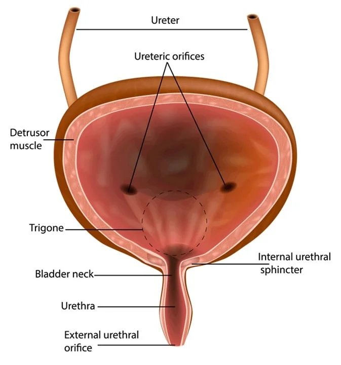
- Muscularis: The middle layer composed of smooth muscle known as the detrusor muscle. This muscle contracts to help expel urine.
- Adventitia: The outer layer that provides structural support and helps anchor the bladder in place.
Bladder Shape and Capacity:
- Empty Bladder: The bladder is small, somewhat spherical, and located low in the pelvis.
- Full Bladder: As it fills with urine, it expands upward into the abdominal cavity and can become more elongated or oval in shape. Its capacity is about 400-450 ml in adults.
Sphincters:
- Internal Urethral Sphincter: Located at the base of the bladder, it is made of smooth muscle and helps prevent the involuntary leakage of urine.
- External Urethral Sphincter: Located further down the urethra, it is made of skeletal muscle and allows for voluntary control over the release of urine.
Ureters and Urethra:
- Ureters: Tubes that transport urine from the kidneys to the bladder.
- Urethra: The duct through which urine exits the bladder and the body. It varies in length and structure between men and women.
Common diseases and problems of urinary bladder
Common diseases of urinary bladder in men and women are as follows:
- Urinary Tract Infection (UTI)
- Bladder Infections (Cystitis)
- Bladder Cancer
- Overactive Bladder
- Interstitial Cystitis
- Bladder Stones
- Urinary Incontinence
- Bladder Prolapse
- Neurogenic Bladder
- Bladder Outlet Obstruction
- Bladder diverticul
- Contracted bladder
- Haemorrhagic cystitis
- Extrophy vesica
Common symptoms of urinary bladder
The symptoms seen in different diseases of the bladder are as follows:
- Frequent Urination
- Urgency to Urinate
- Painful Urination (Dysuria)
- Hematuria (Blood in Urine)
- Incontinence
- Lower Abdominal Pain
- Urine Leakage
- Difficulty Urinating
- Cloudy or Foul-Smelling Urine
- Nighttime Urination (Nocturia)
Diagnostic tests for urinary of urinary bladder
Tests commonly used in the diagnosis of bladder diseases include:
- Urinalysis
- Urine Culture
- Cystoscopy
- Ultrasound
- CT Scan of the Abdomen/Pelvis
- MRI of the Pelvis
- Urodynamics Testing
- Cystogram
- Intravenous Pyelogram (IVP)
- Bladder Biopsy
- Uroflowmetry
Summary
The urinary bladder is a hollow, muscular organ located in the pelvis that stores urine produced by the kidneys. It expands as it fills with urine and contracts to expel it through the urethra during urination. This process helps regulate the body’s fluid balance and remove waste.
Prof. Dr. Emin ÖZBEK
Urologist
Istanbul- TURKIYE

Leave a Reply