
Renal cysts are fluid-filled sacs that form in the kidneys. They can be classified as simple or complex. Renal cysts are usually discovered incidentally during imaging tests such as ultrasounds, CT scans, or MRIs, which are performed for other reasons. Treatment for renal cysts depends on the type and symptoms. If a cyst causes symptoms like pain, infection, or high blood pressure, or if it’s complex and suspicious for cancer, treatment options may include aspiration and sclerotherapy, laparoscopic surgery, or, in rare cases, nephrectomy (removal of part or all of the kidney).
What is renal cyst?
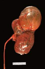
A renal cyst is a fluid-filled sac that forms on or in the kidneys. These cysts can vary in size and number, and they are generally classified into two main types:
Simple Renal Cysts: These are single, thin-walled sacs filled with clear fluid. They are common, especially in older adults. Simple cysts are typically benign and rarely cause complications.
Complex Renal Cysts: These cysts may have septations (internal walls), calcifications, or solid components. They are less common and can vary significantly in appearance and structure. Complex cysts need careful evaluation, as they have a higher risk of being cancerous. Imaging characteristics and other factors are considered to assess their malignancy potential.
Causes of renal cysts?
The causes of renal cysts can vary based on the type of cyst. Here’s a breakdown of potential causes:
Simple Renal Cysts
- Aging: Simple renal cysts are more common in older adults, suggesting that aging plays a significant role in their development.
- Genetic Predisposition: Some people may have a genetic predisposition to developing renal cysts, although this is not as pronounced as in hereditary conditions like polycystic kidney disease.
- Obstruction and Weakness in Tubules: It is thought that simple cysts might form due to the obstruction of the tubules in the kidneys, leading to fluid accumulation. Weakness in the walls of the tubules may also play a role.
Complex Renal Cysts
- Congenital Conditions: Some complex cysts may be present from birth due to developmental anomalies.
- Genetic Factors: Complex cysts can be associated with genetic conditions such as von Hippel-Lindau disease and tuberous sclerosis.
Other Factors
- Kidney Injury or Infection: Past kidney injuries or infections can sometimes lead to the formation of cysts.
- Chronic Kidney Diseases: People with certain chronic kidney diseases are more prone to developing cysts.
- Dialysis: Long-term dialysis can be associated with the development of acquired cystic kidney disease (ACKD), which involves the formation of multiple cysts in the kidneys.
Bosniak classification of renal cysts
The Bosniak classification system is used to categorize renal cysts based on their appearance in imaging studies, particularly CT scans. This classification helps in assessing the likelihood of malignancy and determining the appropriate management. Here’s an overview of the Bosniak classification system:
Bosniak Category I:
- Description: Simple cysts with thin walls, no septations (internal divisions), calcifications, or solid components.
- CT Findings: Homogeneous water density (0-20 Hounsfield units), no enhancement with contrast.
- Malignancy Risk: Almost zero.
- Management: No follow-up needed.
Bosniak Category II:
- Description: Minimally complex cysts with a few thin septations or fine calcifications. Cysts may be slightly hyperdense (< 3 cm) but do not enhance with contrast.
- CT Findings: Thin septations, fine or curvilinear calcifications, homogenous hyperdense cysts.
- Malignancy Risk: Very low (close to zero).
- Management: Typically no follow-up needed.
Bosniak Category IIF:
- Description: Cysts with more septations, minimal thickening of the septa or wall, or thicker, more nodular calcifications. No measurable contrast enhancement.
- CT Findings: Multiple thin septa or minimal enhancement, wall thickening, calcifications.
- Malignancy Risk: Slightly higher, about 5%.
- Management: Requires follow-up imaging to monitor for changes.
Bosniak Category III:
- Description: Indeterminate cystic masses with thickened, irregular walls or septa that may show measurable enhancement after contrast.
- CT Findings: Thick or irregular septa or walls, measurable contrast enhancement.
- Malignancy Risk: Approximately 50%.
- Management: Surgical exploration or biopsy often recommended.
Bosniak Category IV:
- Description: Clearly malignant cystic masses with enhancing soft tissue components independent of the wall or septa.
- CT Findings: Solid enhancing soft tissue components, significant wall or septal thickening.
- Malignancy Risk: Very high (close to 100%).
- Management: Surgical removal is usually indicated.
The Bosniak classification helps guide the management of renal cysts based on their risk of malignancy:
- Category I and II: Typically benign, no routine follow-up.
- Category IIF: Low risk, requires periodic monitoring.
- Category III and IV: Higher risk of malignancy, often require surgical intervention.
Symptoms
Simple renal cysts are often asymptomatic and discovered incidentally during imaging studies conducted for other reasons. However, when symptoms do occur, they can include:
- Pain: Persistent, dull aching pain in the side (flank) or lower back. This is usually due to the cyst enlarging and pressing on nearby organs or tissues. In some cases, pain can extend to the abdomen.
- Hypertension (High Blood Pressure): Elevated blood pressure can sometimes be associated with large cysts, though the exact mechanism is not always clear.
- Hematuria: Visible blood (gross hematuria) or microscopic blood (detected via urine tests) in the urine can occur if the cyst ruptures or if there is bleeding into the cyst.
- Urinary Symptoms: Frequent urination, urgency (a sudden, strong need to urinate) and recurrent UTIs can be associated with renal cysts if they become infected.
- Rare Symptoms: In rare cases, a large cyst may obstruct the flow of urine, leading to hydronephrosis (swelling of the kidney due to urine buildup) or impaired kidney function.
Diagnosis
Here’s a detailed approach to the diagnosis:
1. Medical History and Physical Examination:
- Medical History: The doctor will take a detailed medical history to check for any symptoms such as flank pain, high blood pressure, or urinary issues.
- Physical Examination: A physical exam may reveal tenderness in the area of the kidneys.
2. Imaging Studies: Imaging is the cornerstone of diagnosing simple renal cysts. The following methods are commonly used:
- Ultrasound: Simple cysts appear as anechoic (dark) areas with a smooth, thin wall and no internal echoes.
- Computed Tomography (CT) Scan: Simple cysts show up as round or oval, well-defined areas with fluid density and no enhancement after contrast administration.
- Magnetic Resonance Imaging (MRI): Simple cysts appear as fluid-filled sacs with high signal intensity on T2-weighted images and low signal intensity on T1-weighted images.
3. Bosniak Classification: For evaluating renal cysts, particularly to distinguish simple cysts from complex ones, the Bosniak classification system is often used based on CT findings:
- Bosniak I and II: Typically represent simple cysts that are benign and require no follow-up.
- Bosniak IIF, III, and IV: Indicate increasingly complex cysts that may require closer monitoring or intervention due to a higher risk of malignancy.
4. Laboratory Tests:
- Blood Tests: While not specifically diagnostic for renal cysts, blood tests can help assess kidney function and rule out other potential causes of symptoms.
- Urine Tests: To check for blood or infection that could be associated with kidney issues.
When to see a doctor
You should seek medical attention if you experience:
- Persistent or severe flank or back pain.
- Blood in the urine.
- Signs of a urinary tract infection (e.g., burning sensation during urination, fever, frequent urination).
- Unexplained high blood pressure.
- A noticeable mass or swelling in the abdomen.
Complications
Complications of renal cysts can vary based on their type and size.
Simple Renal Cysts
- Infection: A cyst can become infected, causing fever, pain, and general malaise.
- Rupture: Though rare, a cyst may rupture, leading to sudden, sharp pain in the flank area.
- Hemorrhage: Bleeding into the cyst can occur, resulting in pain and hematuria (blood in the urine).
- Obstruction: A large cyst can obstruct the flow of urine, potentially leading to hydronephrosis (swelling of the kidney due to urine buildup) or urinary tract infections.
Complex Renal Cysts
- Malignancy: Complex cysts have a higher risk of being cancerous, requiring regular monitoring and sometimes surgical intervention.
- Infection and Hemorrhage: Similar to simple cysts, complex cysts can also become infected or bleed.
Indications for treatment
Treatment is usually considered if the cyst:
- Causes persistent pain or discomfort in the flank or back.
- Leads to high blood pressure that cannot be controlled with medication.
- Is associated with recurrent urinary tract infections.
- Shows signs of bleeding, infection, or rupture.
- Grows significantly in size over time.
Treatment
Simple renal cysts are usually benign and asymptomatic, requiring minimal intervention. However, treatment may be necessary if they cause symptoms or complications. Here are the primary treatment options:
Observation: Monitoring: If the cyst is small and asymptomatic, regular monitoring with periodic imaging (ultrasound, CT scan, or MRI) may be recommended to ensure it does not grow or cause problems.
Aspiration and Sclerotherapy:
- Aspiration: A needle is inserted through the skin into the cyst under ultrasound or CT guidance to drain the fluid.
- Sclerotherapy: After aspiration, a sclerosing agent (such as alcohol) is injected into the cyst to help shrink it and prevent it from refilling.
Laparoscopic Surgery: Laparoscopic Cyst Decortication: If the cyst is large, symptomatic, or causing complications, laparoscopic surgery may be performed to remove the cyst wall.
Open Surgery: Open Cyst Decortication: In rare cases where the cyst is very large or there are multiple cysts causing significant issues, open surgery may be required to remove the cysts.
Post-treatment care
- Follow-Up Imaging: Regular follow-up imaging is often recommended to monitor for recurrence or complications after treatment.
- Symptom Management: Pain or discomfort following treatment may be managed with over-the-counter pain relievers or prescribed medications.
Summary
Renal cysts are fluid-filled sacs that develop in or on the kidneys. They can be simple (benign) or complex (potentially malignant). Simple cysts are generally benign and require minimal intervention. Complex cysts may need closer monitoring or surgical treatment due to a higher risk of malignancy. Follow-up is required in asymptomatic cases, and treatment is required if symptomatic and malignancy is suspected. We generally prefer laparoscopic treatment. In this way, full recovery is achieved in patients.
Prof. Dr. Emin ÖZBEK
Urologist
Istanbul-TURKIYE

Leave a Reply